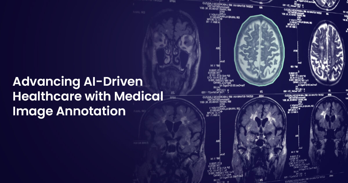
Machine learning is transforming the medical industry, with AI advancing diagnostics and treatment across various specialities. In fields like radiology, AI-powered neural networks assist in image interpretation and disease detection, such as localizing tumors in CT scans. However, AI models are only as reliable as the data they are trained on—biased or inconsistent datasets can compromise performance. Despite the abundance of medical images, the lack of standardized and diverse data, along with privacy concerns, makes implementing AI in healthcare challenging. This highlights the critical role of image labeling in developing accurate and reliable medical AI models.
Labeled data train AI models to recognize patterns in medical images, enhancing diagnostic accuracy and efficiency. For example, AI-powered tools trained on labeled data can efficiently differentiate between benign and malignant tumors in MRI scans, aiding in early cancer detection.
What is Medical Data Image Annotation?
Medical image annotation is the process of labeling and describing medical imaging data, such as X-rays, CT scans, MRIs, ultrasounds, PET scans, and mammograms, to train AI algorithms for image analysis and diagnostics. AI models help doctors save time, make better-informed decisions, and improve patient outcomes.
Medical image annotation enables AI-driven diagnostics across specialties, including radiology, oncology, dentistry, neurology, dermatology, and cardiology. Annotation ensures that AI systems learn from structured and compliant data, leading to accurate and reliable predictions.
Types of Medical Images
Some of the common medical imaging modalities that are annotated include X-rays, CT scans, ultrasounds, MRIs, PET scans, mammograms, echocardiograms, and EEGs. Medical images have specific characteristics that should be considered when they are processed, such as pixel depth, photometric interpretation, metadata, and pixel data.
File Formats for Medical Image Data Storage
Annotated medical images can be stored in different formats depending on the specific use cases and processing requirements. This ensures image quality, compatibility, and effective learning for ML models. Common formats include:
-
DICOM (Digital Imaging and Communications in Medicine): Used for most medical imaging modalities—such as X-rays, ultrasounds, CT scans, MRIs, and PET scans—used in digital radiology.
-
NIfTI (Neuroimaging Informatics Technology Initiative): Primarily used for neuroimaging, including MRI and fMRI scans.
-
TIFF (Tagged Image File Format): Used in research, particularly for storing microscopy images of cells with separate metadata files.
-
WSI (Whole Slide Image): Suitable for digital pathology, storing high-resolution histological (microscopic) images.
-
JPEG (Joint Photographic Experts Group): Used as a compression method within DICOM files, such as compressing CT scan images.
-
BMP (Bitmap Image File): Commonly used in research, typically alongside metadata files in plain text or standard formats, for example, storing digitized photographs of skin lesions.
-
PNG (Portable Network Graphics): Suitable for research, typically with separate metadata files, such as storing diagrams of anatomical structures.
Dimensions of Medical Images
-
2D images: Generated by X-ray, ultrasound, or electroencephalography.
-
3D images: Generated by CT scans and MRI scans.
-
4D images: Generated by dynamic cardiac MRI and dynamic PET scans.
Types of Medical Image Labeling Tasks
-
Image Classification: A fundamental annotation technique that involves dividing medical images in categories based on specific attributes, such as identifying pathologies (e.g. tumors) within medical images or analyzing disease severity.
-
Object Detection: Focuses on precisely identifying and outlining specific anomalies, such as tumors, lesions, and other abnormalities, or anatomical structures, such as organs and bones. This aids in identifying critical regions of interest.
-
Semantic Segmentation: A pixel-level annotation method where each pixel is assigned a label, effectively creating a color-coded map of different tissue categories with an image. This level of detail is extremely helpful for applications like brain imaging and cancer treatment where differentiating healthy tissues from cancerous ones is pivotal for treatment planning.
-
Landmark Annotation: Involves marking key points within an image for accurate measurements and analysis, which is used in facial recognition, surgical planning, and post-surgical evaluations.
Use Cases
The application of annotated medical images goes beyond disease diagnosis and detection. It serves various purposes across multiple specialities, redefining how healthcare is delivered and advanced.
-
Disease Diagnosis and Detection: Imaging annotation enables accurate and streamlined disease diagnosis by marking abnormalities such as tumors, lung nodules, or other pathologies in medical images. This facilitates early disease detection.
-
AI/ ML Model Training: AI and ML models trained on annotated data can detect diseases, identify anomalies, and assist in diagnosis. Diverse, compliant and accurate data enhances algorithm performance and clinical decision-making.
-
Treatment Planning and Monitoring: Annotation aids in surgical planning, ensuring precision and minimizing risks. Follow-up images help monitor patient progress post-treatment.
-
Medical Education and Training: Annotated images provide a clear and comprehensive visual representation of cases, helping medical students and practitioners get deeper insights into anatomy, disease, and treatments.
-
Drug Development and Clinical Trials: Annotation helps measure disease progression, such as tracking tumor size changes. The precise measurement allows researchers to determine the efficacy of new drugs or therapies.
Challenges in Medical Image Annotation
Annotating medical images comes with several challenges, largely due to the complexity of different imaging technologies.
-
Low Contrast Between Tissue Types: In radiology, low contrast between different tissue types makes it difficult to separate organs and structures in X-ray, CT scan, and MRI images. This makes it challenging to determine the ‘ground truth’, especially for complex cases like tumor identification.
-
Multidimensional Data: Many medical images, such as CT scans and MRIs are often captured as a series of slices, forming a 3D volume, which require 3D annotation with specialized tools and expertise.
-
Unique File Formats: Radiology images come in specialized formats like DICOM or NIfTI, which are not supported by some data labeling tools. Moreover, 4D cardiac CT scans pose processing challenges.
-
Lack of Data Diversity: Obtaining diverse and consistent medical images, especially of healthy patients, is challenging due to privacy and security laws. Public datasets may lack reliability for training AI models.
-
Domain Expertise: Medical images require specialized knowledge for accurate interpretation. This necessitates employing healthcare professionals like radiologists, physicians, and even surgeons, which is costly and time-consuming.
-
Time-Consuming Process: Annotating medical images requires human annotators to draw outlines around anatomical structures and anomalies. Drawing and editing polygon masks is a labor-intensive task and consumes a lot of time, especially for 3D images.
-
Data Anonymization and Compliance: Medial data contains sensitive patient information. DICOM files, for example, often contain personally identifiable information (PII), which must be anonymized. Annotation processes must follow strict privacy regulations like HIPAA.
Best Practices for Medical Data Annotation
Here are some of the best medical image data annotation practices to produce quality training datasets.
-
Multidisciplinary Team: Having board-certified medical professionals and annotators from diverse geographies helps reduce individual bias and improve reliability.
-
Diversity in Data: AI training data should represent different demographics, conditions, and imaging variations to improve generalization and prevent bias.
-
Standardized Labeling Guidelines: Following standardized guidelines ensures consistency and makes data suitable for clinical and AI research.
-
Automation: AI-enabled tools can be used to speed up the annotation process and reduce human effort where applicable.
-
Quality Auditing: Senior healthcare professionals such as radiologists, physicians, and surgeons should double-check annotations to verify accuracy and consistency.
-
Regulatory Compliance: Annotation processes must comply with data privacy and security regulations such as HIPAA, FDA, and GDPR to protect patient information.
Final Thoughts
Annotating medical imaging is fundamentally different from regular data annotation due to complexity, specialized formats, expert annotators, and strict regulatory requirements. These images use unique file formats like DICOM and NIfTI, multiple slices and views, and radiology-guided controls to ensure consistency across volumetric data.
By combining advanced annotation technologies, multiple expert reviews, and standardized protocols and regulations, high-quality imaging data can be created to power reliable medical AI models for various applications.


