
Osteogenesis Imperfecta: Understanding the brittle bone disease
1. Introduction
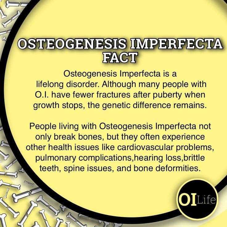
Miscellaneous diseases may disturb the equilibrium of bone metabolism. Any alteration in bone remodeling, deficiency of parathyroid hormone or/and active vitamin D1-hydroxylation may lead to significant changes in the geometry or structure of the bones. There are also various known diseases in the bone tissue that can cause hypercalcemia and hypocalcemia. Osteogenesis Imperfecta (OI) or ‘Brittle Bone Disease’, a rare genetic disorder, may be one of those diseases that may eventually cause serious changes in a patient’s skeletal mass. OI is characterized by fragility and continuous fractures in the bone that occur after minimal trauma. Although the majority of cases are sporadic with a typical complex infrequent pattern, they are inherited as autosomal dominant or recessive. Additionally, to fractures and bone disorders, there could be a broad range of symptoms and situations related to OI including blue sclera, opalescent teeth, reduced height, hyperlaxity, macrocephaly, malocclusion, anodontia, hydrodentia, altered ENT, an altered cardiovascular system, and some of the neurological difficulties among other points related to the general welfare of the patient.
As we are exposed to various diseases and/or diseases, the continuous weaving and unraveling of bone tissue is necessary not only to maintain their integrity but also to support their metabolic functions, such as sodium, calcium, and phosphate storage and hormonal balance. This process is structured in what is known as ‘bone remodeling’, a process common to all bone tissue that continually removes and creates new tissue. When remodeling, each part of the bone is done in both an independent and coordinated manner. Sometimes, during inappropriate remodeling activity, diseases arise. The present article aims to clarify such a process and the risks and challenges that are faced when such events arise during bone metabolism.
Bone is a living tissue that is constantly being remodeled and changed. Throughout life, bone tissue is renewed as old bone is resorbed and new bone is formed. This process can be influenced by hormones, such as those associated with an individual’s growth and overall health. Some of the hormones that may affect this process are parathyroid hormone and the active hormonal form of vitamin D, which are essential to overall bone health. For instance, when blood levels decrease, bones release stored calcium and phosphate into the bloodstream in a process called resorption.
2. Causes and Risk Factors
The risk factors for OI are parental consanguinity, a de novo genetic change that is not present in either parent (approximately 15–25% of cases), high rate of recurrence, and multisystem involvement. The risk of having a child with the same clinical phenotype is approximately 50% and depends on the genotype, allelic heterogeneity, and effects of rare polymorphisms, molecular modifier mechanisms, genetic, epigenetic, and environmental factors, such as temperature, hypothesis of the founder effect, and epigenetic imprinting of phenotypic variability for AD forms. At the same time, there is no risk of having an affected child if the parents and the affected child have “negative” genetic testing, and the structure of the parents’ chromosomes is normal. The P3H1 and CRTAP gene alterations in OI also affect other non-collagenous extracellular matrices, retroviral surface receptors, certain bacteria, and enveloped viruses. Different signaling functions can contextually modify the OI phenotype produced by glycine substitutions and their impact on collagen deposition and other ECM-dependent cell functions, including differentiation, proliferation, migration, and survival. Neural expression is sensitive to both decreased EXP1 and increased GFR expression levels, which can enhance adhesion and proliferation signaling. Neural cell response to proper ECM processing provided by P3H1 and CRTAP is by increased collagen deposition and expression of appropriate alpha1P and alpha2P bonding motifs usually produced by processing vitamin C-dependent P3H1 hydroxylases.
There are two main causes of osteogenesis imperfecta (OI): dominant and recessive mutations in the COL1A1, COL1A2, CRTAP, LEPRE1, PPIB, FKBP10, SERPINF1, TMEM38B, WNT1, CREB3L1, LEPR, SP7, and BMP1 genes, which are inherited in an autosomal dominant (AD), autosomal recessive (AR), or X-chromosome linked form. COL1A1 and COL1A2 encode the Proα1 and Proα2 chains of collagen type I, respectively. Mutations in these genes can lead to defective production of the Proα1(I) or Proα2(I) chains or abnormal sequences within these chains, preventing correct formation of the collagen helix. These are the typical dominant (haploinsufficient) mutations found in the AD form of OI. They can also cause predominantly qualitative defects affecting the ability of one-third of collagen helices to incorporate the mutation with subsequent formation of pathologically altered collagen fibrils. CRTAP, P3H1, and cyclophilin B also play an important role in the posttranslational modification and structure of the collagen molecule by facilitating the isomerization of prolines (P) present in the Gly-X-Y sequence of collagen type I chains and can be associated with the AR forms of OI. While CRTAP and P3H1 form a complex with cartilage-associated protein cyclophilin B (CYPB), their combined effect has been shown to control the proper folding of the triple helix and the rate of correct folding and assembly of the molecule into the triple helices and productive overmodification of collagen type I. Disorders of collagen I and structure lead to dominant and recessive forms of OI. Depending on the severity and type of mutation, you can get a different expression and OI phenotypes.
3. Symptoms and Diagnosis
Osteogenesis imperfecta is usually attempted to be diagnosed prenatally. This might happen during routine ultrasonography, or CAP-auscultation of Amniotic Fluid and echocardiography are further ratio-chinese tests for parents of the patients carrying a pathogenic mutation causing during the genes that encode I lost its _development and re-formation. The syndrome may be immediately genetic counseling. Further genetic tests may be accomplished as early as the 7th-8th week of gestation, mapping out to non-invasive prenatal check outperforms own over the traditional strategies, such as from on a sex chromosomes. Implantation of either sex-imposed intrauterine means, by which biopsies are conducted from the embryonic cells or the placenta. More lately, Chorionic Villus Sampling mess-ups utilizing adherent mothers’ cells may come across our stop, not support the initiative from this area. If prenatal assays are not practiced, postnatal medical diagnosis can become a great deal made equally by c-FLaSK tests (Combination of Ligation specific PCR and adapter reliant split circle ligation) done as early as 42 days. Peripheral Blood, umbilical cord blood, or perhaps skin fibroblasts can be examined. Sealing the definitive diagnosis normally requires 2 to three weeks.
This bone is labeled as “brittle bones”. Due to the different mutation types and location in the DNA, there is an expansive range of the severity of this syndrome, amongst which patients with type I and type IV are often encountered. We will split this extensive range into different types of syndrome. Moreover, we will inform about the possible underlying mechanisms and focus on the signs of the syndrome being reflected in the eye. Until now, there is no effective treatment available, however, stem cells do represent a promising form of therapy.
4. Treatment Options
Medical management will include the use of surgical techniques to correct alignment of the long bones as needed, decreasing bone pain as much as possible, and stabilizing fractures early to prevent bone deformities. For those with moderate and severe OI types, medical management will include different drug approaches to help increase bone density and decrease fracture rates. There are different “bisphosphonate” drugs that can be used to treat OI, and more trials of different drugs are ongoing. Other new therapies for the two common severe OI types are also being tested, which may help to increase the amount of good bone in these types of OI.
Even though there is no cure for OI, many of the problems associated with OI can be treated and managed. In addition to working with a variety of medical specialists, families will also need to learn how to best care for and help support the child. Medical management and working with a variety of medical specialists may help to keep the complications of OI to a minimum or decrease the severity of these complications.
Bone Mass Measurement in Osteogenesis Imperfecta: The Osteogenesis Imperfecta Foundation and The Osteoporosis Foundation sponsored a workshop to develop guidelines for bone mass measurement in OI. The following guidelines, developed from the workshop statements, are provided with commentary and literature review. The resulting guidelines do not supersede individual professional judgment in patient evaluation or practice and are meant to be used as a tool to determine necessary exams to be obtained in OI.
Emerging Therapies: A Report from the Osteogenesis Imperfecta Foundation’s Workshop on Treatment of Osteogenesis Imperfecta discussed emerging therapies such as samarium and growth hormone. Bisphosphonates continue to be the recommended treatment for moderate OI types, and other treatments should be done in well-organized clinical studies to show the benefits of other OI treatments.
5. Living with Osteogenesis Imperfecta
In the United States, there are two active national organizations for people affected by OI, the Osteogenesis Imperfecta Foundation and the OI Foundation. Both groups play an active role in a number of different topics, such as research, and can assist you with networking and finding resources. The YODA project (Your OI Data Access), hosted by the Osteogenesis Imperfecta Foundation, serves to collect medical data from people living with OI for analysis by researchers. With only two exceptions, no data entered into the YODA Project’s database is kept with other patient identifiable information. The data entered by patients is kept confidential and cannot be used to identify individuals.
Children with OI have several healthcare needs. How much medical care a child with OI requires, and which doctors provide that care, varies depending on the child and the severity of his or her OI. Many, but certainly not all, children with OI receive care from a bone specialist or orthopedist. Other common medical specialists include geneticists (a doctor who studies inherited conditions) and ophthalmologists (a doctor who studies the eye). The care of a child with OI is usually coordinated by a pediatrician or a family practice doctor. In addition, any child with OI should have an adult dentist, as well as a pediatric dentist, and families are often surprised to learn that the old recommendation that children with OI be on a “soft diet” might not be the best way to ensure good dental health. Many young adults receive their care from a doctor who treats adults; such a physician might be a general practitioner (a doctor, also referred to as a primary care provider, who can provide routine, everyday healthcare), a family practice doctor, or a general internist (a doctor who offers comprehensive care for adults).